Description
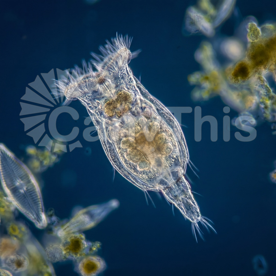
Rotifera — microscopic animals found in all freshwater environments — are among the most complex organisms visible at the optical microscopy scale, their 1,000-cell body containing differentiated organ systems including a brain, eyespot, stomach, and the signature rotating corona of cilia that give them their name. This photograph was made using a compound microscope with a photomicroscopy-configured camera and Köhler illumination at 40:1 optical magnification, the organism maintained in a thin, controlled-depth water layer. The corona cilia creating the rotational water current, the transparent body allowing visualization of the internal mastax jaw structure, and the eyespot are all visible in a single frame of exceptional scientific documentation value.