Description
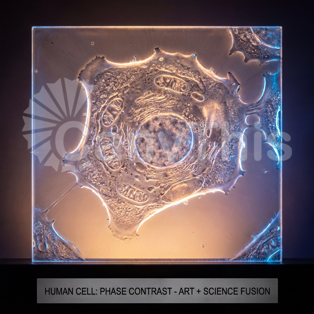
Phase contrast optical microscopy allows transparent biological specimens to be imaged without staining — the refractive index differences between different cellular components produce phase shifts in the transmitted light that the phase contrast optical system converts into amplitude variations (bright and dark regions). At 40× magnification, a human cell in metaphase — the stage of mitosis when chromosomes are aligned at the cell’s equatorial plane — shows the condensed chromosomes as dark, high-contrast structures approximately 2-5 microns in width, clearly distinguished from the surrounding cytoplasm. This image captures two adjacent cells in different mitotic stages: the cell on the left in metaphase (chromosomes aligned at the metaphase plate), and the cell on the right in anaphase (chromosomes separating toward the cell poles). The non-dividing cells surrounding the dividing cells are visible in their interphase morphology — irregular and flat with a large, pale nucleus.