Description
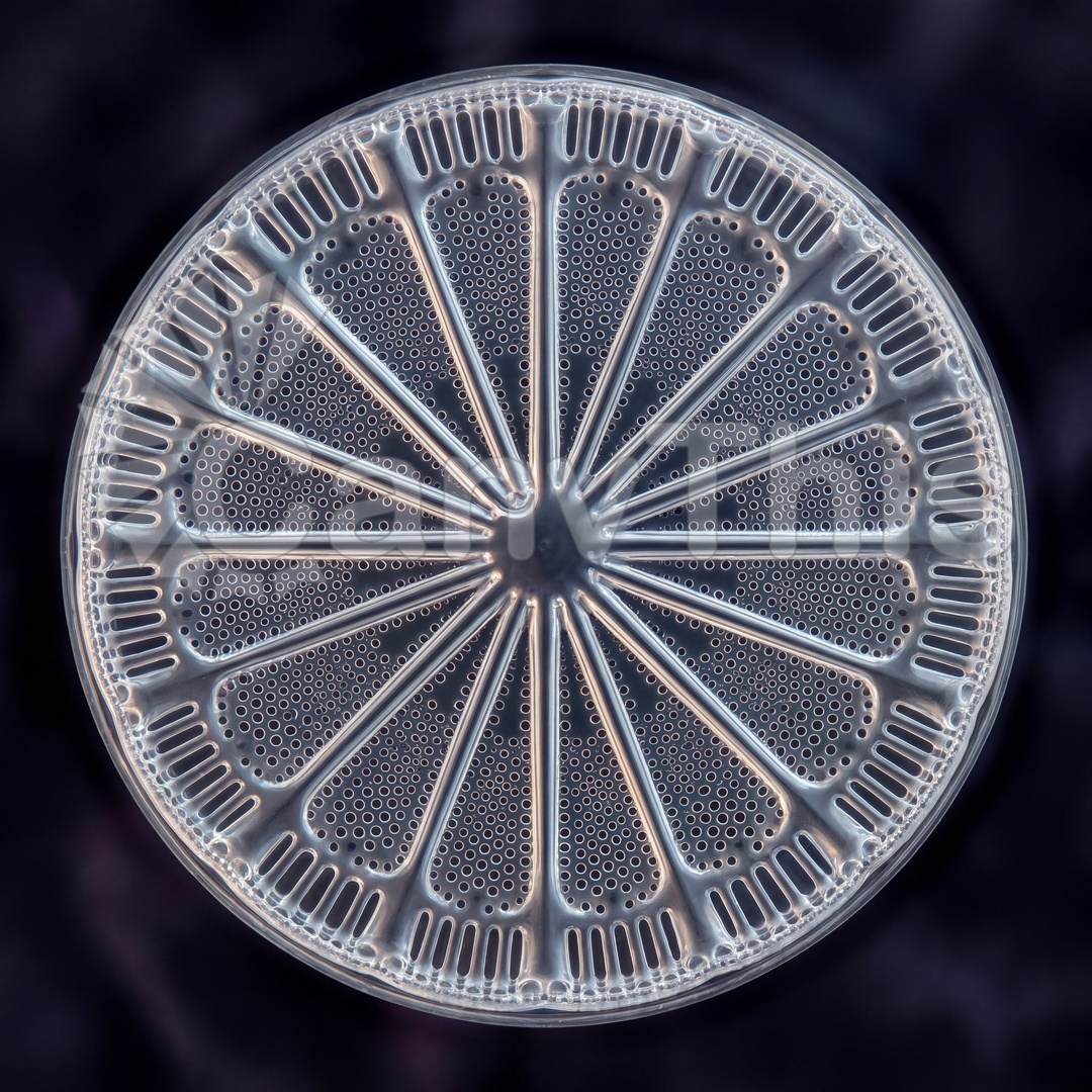
Diatom frustule photography using dark-field illumination at optical microscopy magnification produces images of extraordinary delicacy — the silica shell of these single-celled algae, with its regular perforation arrays, diffracts the dark-field illumination into vivid colour fringes that resolve the shell’s three-dimensional architecture in full colour without the vacuum environment of electron microscopy. This image was made using a research-grade optical microscope with dark-field condenser at 40× objective, the diatom sample prepared from a freshwater pond sample concentrated by centrifugation. The species shown — a pennate diatom of the Pinnularia genus — shows the characteristic bilateral symmetry of the pennate form, its raphe groove running the full length of the frustule, and the regular pore array of the valve face. The dark-field illumination renders the frustule as a glowing structure of golden light against the black background.