Description
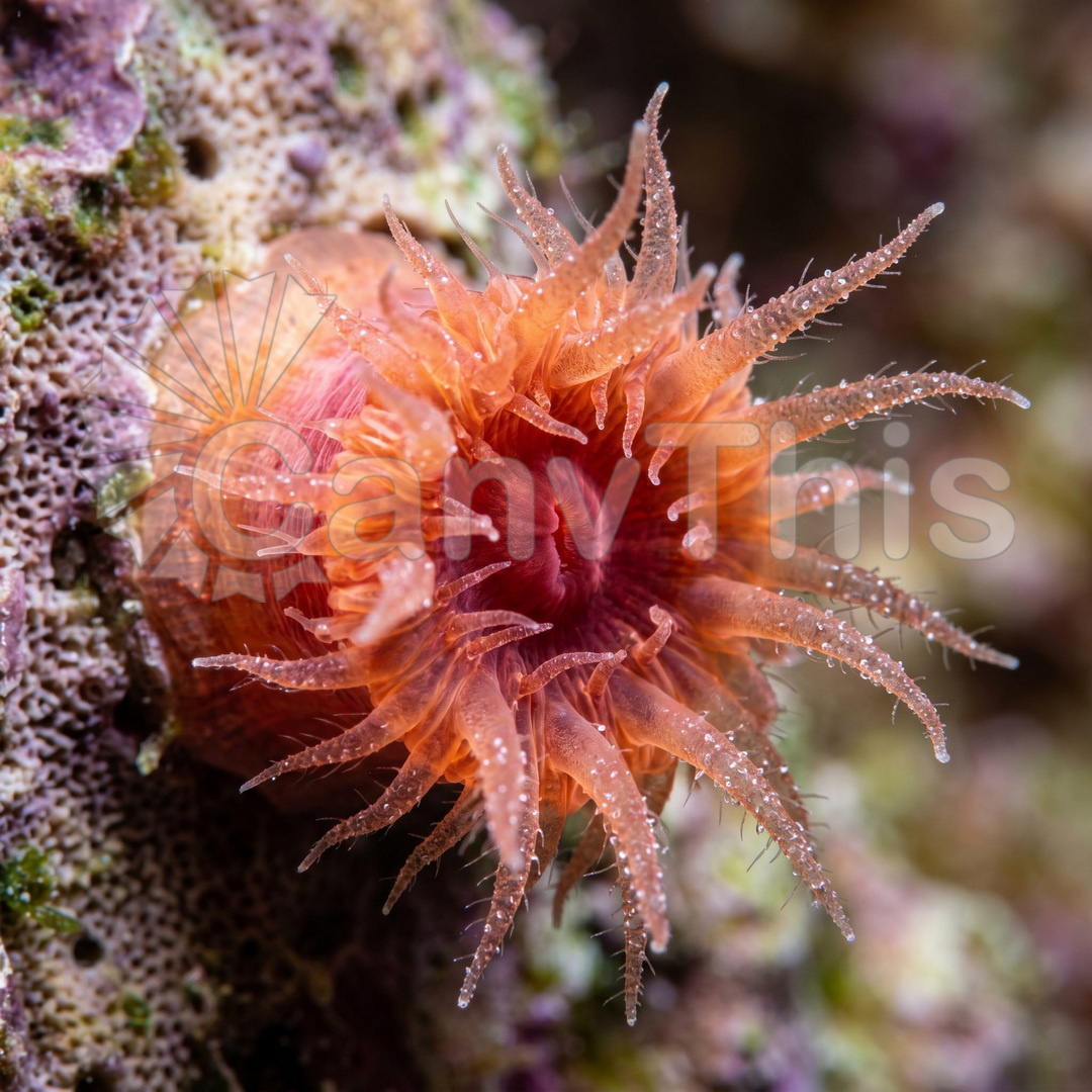
Coral polyp photography at 5:1 macro requires either night diving conditions (when polyps extend their tentacles for feeding) or aquarium subjects. This image was made of an aquarium-maintained brain coral Diploria specimen under controlled lighting conditions, the polyp extending its tentacle crown as the water flow was reduced to simulate the low-current nighttime conditions that trigger feeding extension. The full tentacle crown of a single polyp is visible at 5:1 reproduction: 24 tentacles arranged in two concentric rings around the central mouth disc, each tentacle approximately 3mm in length and bearing the distinctive spherical nematocyst batteries at its tip. The nematocysts — the stinging organelles that paralyse zooplankton prey — appear as the slightly darker, denser tip regions of each tentacle. The skeletal calyx walls of the coral skeleton are visible below the polyp as the white calcium carbonate structure that the polyp secretes during its upward growth.